


Lumbar osteochondrosis is a disease that deforms and destroys the cartilaginous tissue of intervertebral discs in the lower back.With no layer of cartilage, the distance between the vertebrae is significantly reduced.And with the smallest sharp curves, they can change.The main danger of the disease is the possibility of intervertebral hernia formation.
Can't you lean to create an object that has fallen to the floor?Do you suffer acute pain in the lumbar spine and usually go, involving the waist in a hot scarf?Do not ignore the condition that bothers you.
Lumbar osteochondrosis can drag itself for a long time.There is no need to try the body by force.Love your body.And that will return.
The lumbar region is responsible for most of the load of all body weight compared to the chest and cervical departments.Therefore, these subspecies of osteochondrosis are the most common.
What are the stages of the development of osteochondrosis?
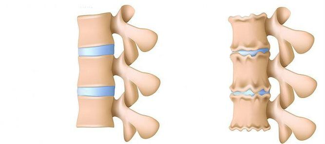
- 1st stage.Prevented.The height of the disk is reduced.In the fibrous ring (the outer layer of the intervertebral disc of cartilaginous fibers) a crack is formed.Lumbar muscles begin to get tired quickly.You feel some discomfort on your back.
- Stage 2. Violations of metabolic processes in the core jacket (central part of the intervertebral disc, which consists of a cartilage jacket): their cells are dead or completely destroyed.The structure of the collagen (the protein structure is based on the connective tissue) of the fibrous ring is also disturbed.Local pain, a person cannot deal with the physical activity that previously considered quite viable.
- Stage 3. Full destruction of the fibrous ring.Adjacent vertebrae are no longer stable.Any uncomfortable pose causes pain.Due to the experience of nerve roots that move away from the spinal cord, limbs can become less sensitive and mobile.
- 4th stage.Intervertebral disk tissues become scar.The vertebra may be in the shell.The clinical description here depends on individual physiology.
Lumbar pain (lumbago) and the pain that gives to the leg during the sciatic nerve (Ishias) is one of the most common complaints that patients seek medical help.Due to the fact that these symptoms are quite common in the general population, and their constant growth is also observed, the diagnosis and treatment of these patients will remain one of the main areas of activity of neurosurgical hospitals.Despite the generalization of this pathology, surgical removal of intervertebral disc hernia (MPD) is required only in 10% of patients with the clinical picture of the lumbar algae.In the remaining part of the patients, the best effect has conservative treatment, including drug therapy, physical therapy exercises, the use of treatment physiotherapy methods, as well as a return to anterior daily physical activity.
Disease stages
Degenerative-distribution processes usually begin with a deterioration in the intervertebral disc shock absorption function.
- Deterioration of blood supply to the intervertebral disc.In adults, the food of intervertebral discs is performed by diffusion: blood is delivered only to vertebrae and already through them “infiltrates” the discs.In the best way, the disk is fed during dynamic loads (for example, walk), as the pump principle (processed fluid output when tablet, nutrient flow and oxygen when removing the load).Thus, the nutrition of intervertebral discs is difficult, especially under the conditions of a sedentary lifestyle (hypodynamamia).
- Changes in the core of the disk pulpic.With a deterioration in the blood supply, the supply of water, sugars and amino acids in the core of the pulpae is disturbed.For this reason, the production of carbohydrates that connect water suffers.The nucleus is dehydrated, its structure made of gel in a fibrous gel, the ability to drag and extinguish the shots worsen.This increases the load in the fibrous ring and vertebrae, it is more likely to be blocked and injured.
- Changes in the fibrous ring of the intervertebral disc.Due to the flattening of the pulpee nucleus, the increase in the load is in the fibrous disc ring.Under conditions of bad blood supply, the fibrous ring loses its strength.The instability of the spine occurs, which can lead to the formation of an intervertebral hernia, a displacement of vertebrae and damage to the spinal cord or nerve roots.
- Disk protrusion.The formation of intervertebral hernia.As fibrous ring fibers weaken, the Pulpic nucleus begins to stand out, for example, towards the intervertebral channel (disk protrusion).This impressive can also lead to a rupture of a fibrous ring and the formation of a hernia.Read more about the process of intervertebral hernia formation in a separate article - "effective treatment of intervertebral hernia at home."
- Spondylosis is the destruction of intervertebral joints (spondylartrosis), osteophyte growth and ligament ossification.Parallel to intervertebral hernia formation in osteochondrosis, intervertebral joint damage, destructive changes in the vertebra (cartilage) and ligaments are observed.
As osteochondrosis and the development of complications progress, you need to resort to medication increasingly increasing the doses.This leads to high financial costs as well as additional health deterioration due to the side effects of drugs.
Drug therapy, as a rule, is complemented by the immobilization of one or friend of the spine using orthopedic score of varying degrees of rigidity.
Surgical treatment is justified only in cases where the level of compression of the spinal tower, determined by the clinically, corresponds to the exam confirming the rupture of the fibrous ring with the "loss" of MPD hernia in the lumen of the vertebral canal [3-6].The results of surgical treatment in patients with small disk protrusions are usually disappointed with doctors and the patient himself.The method for establishing an accurate diagnosis is magnetic resonance imaging (MRI).Approximately 10% of people in a common population are impossible to perform routine magnetic resonance imaging because of claustrophobia (fear of closed spaces).In this category of people, it is possible to use the "open" magnetic resonance imaging, with the corresponding loss of the quality of the images obtained.Patients who have already undergone surgical treatment should perform a contrasting magnetic resonance imaging to delimit the post -operative changes of the scar -decay of the true hernial protrusion of the disc.In patients with suspected MPD's hernal protrusion, when the implementation of magnetic resonance imaging is impossible, or the results obtained are misinformation, computed myelography (TC) acquires a special diagnostic value.
Private diagnosis experts who interpret studies, as a rule, exaggerate the degree of disc damage due to the impossibility of comparing clinical data with "findings" during tomography.Conclusions such as “changes correspond to the patient's age” are almost never found in research protocols.Despite the improvement of neuroimaging techniques, the responsibility for the correctly deceived diagnosis is on the clinician's shoulders, as only he can compare the clinical picture with the data obtained during the tomography.An increase in the resolution of somewhat improved improved tomography of surgical treatment, but the deviations of the standard in asymptomatic patients began to be detected.The process of processes that accompanies the degenerative -Distortical lesion of the column has undergone serious progress in recent years.Arthropathy of the arched joints is widespread in the general population and is detected quite oftenIn people of the average and older age group during the MPD degenerative CT research, which are also widely used, they are often detected and MRI is a more specific method for diagnosis.At the same time, the changes pronounced in the MPD are not uncommon, not accompanied by a fibrous ring rupture, but only manifested by a slight "stab" of the spinal canal lumen or intervertebral holes.In some cases, degenerative processes that occur in MPD may lead to the destruction of the fibrous ring with subsequent ruptures, which causes migration of part of the pulpitic core off the disc with compression of the adjacent roots of the spinal cord.The statement that if leg pain is observed, it must necessarily be violated in the roots of the spinal cord is not totally true.For buttock pain with irradiation on the posterior surface of the thigh, it can lead to the degeneration of the MPD itself and the arched intervertebral joints.For a true attack of ishialgia caused by Koreshka compression of the MPD hernia nerve, pain radiates the posterior surface of the thigh and leg.A indefinite pain, limited only to the gluteal area or the area of the thigh without distribution along the sciatic nerve, as well as bilateral pain in the gluteal or hips that change its location (right and left), are caused more frequently by joint arthropathy or diffuse MPD degeneration.The clinical image of Koruska compression of MPD hernia can also be a concomitant pathology (eg knee joint arthrosis).In patients with such pain, surgical treatment will not have the proper effect, regardless of which pathology will be detected by tomographic examination.In other words, in patients with the back pain clinic only, the removal of MPD hernia will be ineffective, even if the tomograms are determined by the MPD protrusion, such as usual and happen.But there are also patients in which the typical image of Ishias is accompanied by a pronounced deactivated pain syndrome, while during studies done using highly perceptive tomography, compression of spinal cord roots is not determined.This category of patients is inadequate to perform surgical intervention, because over time, root symptoms, as a rule, decrease.
It is necessary to clearly imagine the mechanisms that lead to the development of the MPD hernial protrusion, in order to recommend to patients the volume of allowed movements, without forgetting about the work activity.Forces that contribute to the formation of hernal protrusion are the result of degenerative changes in the MPD and a diminishing vertical (height) of the fibrous ring and the core of the pulpae.The MPD stab fragment by 80% changes in the backward direction -while introducing the spinal canal lumen and the medial sections of the intervertebral orifice.This displacement of MPD hernia towards the middle line is facilitated by the retention force of the posterior longitudinal ligament.Up to 10% of hernial protrusions are located laterally and spread to the intervertebral hole (forsin hernias) or on the outer edge of the hole where the cerebrospinal column comes out of it, squeeze it like that.
In the vital activity process, dehydration and degenerative changes lead to the loss of MPD height.These pathological processes involve a fibrous ring and a pulpitic nucleus.The most pronounced destruction of the pulpe core in the backdrop of the concomitant degeneration of the fibrous ring, as a rule, leads only to the loss of MPD height without its significant meetings.With predominant changes in the fibrous ring, the vertical forces that affect the preserved Pulpic nucleus that are a derivative of their own weight, as well as the back muscles, acting on the disc in the side, exert excess pressure on the remaining fragment of the pulse nucleus, which is not able to react the fibrous ring.
The sum of these two forces leads to an increase in centrifugal pressure in the MPD, which, together with the stretching component that acts on the fibrous ring fiber, can lead to its rupture and fragment of fragments of the remaining pulp nucleus.After a hernal protrusion was formed, and the "redundant" fragment of the pulpitic nucleus was outside the fibrous ring, the MPD structure becomes stable again [2].As a result of the forces that affect the degeneratively altered core and the MPD's fibrous ring, they are balanced and its vector, which contributes to the additional protrusion of core fragments, disappears.In some cases, partial degenerative changes in the nucleus Pulpos contribute to the formation of gas within the MPD, followed by excessive pressure on its remaining fragment.The formation of a hernia is also accompanied by the process of gas formation within the disc.
Excessive and clear physical activity shown on the patient's back against the backdrop of the existing degenerative lesion of the spine is usually just a trigger that leads to a detailed clinical image of a compression root syndrome, which is often and erroneously considered by the patients themselves, such as the primordial of lumbaria.Clinically, MPD hernia can manifest with reflex and compression syndromes.Syndromes are referred to to compression, in which the above hernral protrusion is pulled, squeezed and deformed, the blood vessels or spinal cord are compacted and deformed.Reflex reflexes include syndromes caused by the effects of disc hernia on the receptors of these structures, especially the end of the spinal nerves of return, which leads to the development of reflex disorders and tonic manifested by vasomotor, dystrophic and myopascial disorders.
As noted above, surgical treatment with degenerative -discrete from Posvinor is advisable only in 10% of patients, the remaining 90% react well to conservative measures.The basic principles of using the last are:
- Relief of pain syndrome;
- Restoration of the correct posture to maintain the MPD fixation capacity changed;
- Elimination of muscle and tonic disorders;
- restoration of blood circulation in roots and spinal cord;
- Normalization of conductivity in nerve fiber;
- elimination of scarred changes and spacing;
- Realocation of psychomatic disorders.
Treatment
Today, in the treatment of osteochondrosis and its complications, medicines of the following groups are used:
- NET -Anti -Liquid Inflammatory Drugs (NSAIDs) -In the form of tablets or injections of medicines.These funds have the ability to reduce pain, reduce inflammation activity.However, the effect of its use does not last long - from several hours to two to three days.Therefore, these funds should be received for a long time - weeks and sometimes months.At the same time, these medications negatively affect mucous gastrointestinal membranes.Its long -term reception is full of the development of gastritis, ulcerative lesions.In addition, they can negatively affect the work of kidneys, liver and contribute to the development of hypertension.And at the same time, these funds do not contribute to the cleaning of dead cell discs.Therefore, its use is only a way to relieve symptoms for a while, but not eliminate the main problem.
- CTOID Anti -inflammatory drugs (gopmonal).As a rule, they are used for serious and impenetrable pain that accompany hernia, radiculitis, ishias, etc.Gopmons have the ability to eliminate inflammation manifestations (due to the oppression of the immune system), alleviate pain.But they also negatively affect the mucous membranes of the stomach and intestine, promote bone calcium leaching, inhibit the production of their own gopmons.And do not contribute to cleaning the focus of dead cells.
- Popesmolic are drugs that affect muscles or nerves that go to the muscles and cause relaxation of skeletal muscles.This means helping to relieve muscle staples for a while, reduce pain and improve blood flow.But at the same time, they do not help clean the tissue of the dead cells.Therefore, they do not contribute to cure osteochondrosis.
- Epiduppal block - The introduction of painkillers and single agents into the space between the solid brain shell and the periosteum that covers the vertebrae.It is used, as a rule, for intense pain - in the acute period of intervertebral hernia, with severe radiculitis, Ishias.Depending on the composition, this injection helps relieve pain for a period of several hours to several days.After the expiration date, the manifestations of the disease are returned, because the procedure does not help restore metabolic processes on discs.Also, when performed, there is a risk of injury to blood vessels and nerves.
Conservative treatment methods include various orthopedic effects on the spine (corset immobilization, traction, manual therapy), physiotherapy (therapeutic massage, physiotherapy exercises, acupuncture, electrotherapy, mud, various types of heating), paravertebral, peridural block and drug tension.The treatment of degenerative -Distrophic lesion of the spine should be complex and phased.As a rule, the general principle of conservative measures is the appointment of painkillers, non -esteroid anti -inflammatory drugs (NSAIDs), muscle relaxants and physiotherapy.
The analgesic effect is achieved by the appointment of diclofenac, ketoprofen, lornoxicam, tramadol.An analgesic and anti -inflammatory effect pronounced has loroxes, existing in injection and tablet forms.
NSAIDs are the most commonly used drugs for degenerative damage -distribution in the spine.They have an anti -inflammatory, analgesic and antipyretic effect associated with the suppression of cyclooxygenase enzyme (COC -1 and TSOs -2), which regulates the transformation of arachidonic acid into prostaglandins, prostacy, thromboxans.In the elderly and patients with risk factors for side effects, it is advisable to "cove" gastrothetic "under the coverage.In these patients, after the conclusion of the course of NSAID injection therapy, the transition to the compressed forms of the co -2 inhibitors, which have less severity of the side effects of the gastrointestinal tract, is advisable.
To eliminate pain associated with increased muscle tone, it is advisable to include central muscles in complex therapy.
Surgical treatment of degenerative lesion -District of the spine is justified with the ineffectiveness of complex conservative measures (within 2-3 weeks) in patients with MPD hernias (usually more than 10 mm) and non -awaiting root symptoms.There are emergency indications for surgical intervention with a "discarded" sequence in the lumen of the spinal canal and express compression of spinal cord roots.Development of caudal syndrome is facilitated by acute radiculomilohemia, leading to severe hyperalgic syndrome, when even the prescription of drug painkillers, the use of blockade (with glucocorticoid and anesthetic) does not reduce pain severity.It is important to note that the absolute size of disc herniated has no determining value to make the final decision on surgical intervention and should be considered in connection with the clinical condition and findings detected by the tomographic examination.In 95% of cases, open access to the vertebral canal is used in hernia.Several disagreement techniques (cold coagulation, laser reconstruction, etc.) have not been today, and their use is justified only for MPD protrusions.Classic micro -arcure removal of disc herniated is performed using microcurgical tools, binocular extensions or an operational microscope.Analysis of the results of distant treatment (in more than 2 years) 13,359 patients undergoing MPD hernia removal, 6135 of which the kidnapping was removed and 7224 aggressive discsctomy was performed, showed that the relapse was found in which pain was found in terms of pain, which was not found in which the relapse was found than the relapse was found in a little more thanRelac was found in a little more than the relapse was found in the relapse that the relapse was found, which was found 2.5 times more (27.8.8% compared to 11.6%) in which the relapse was found in the relapse that the relapse was found in terms of pain.Probably (7% versus 3.5%) in patients who were only removing the kidnapping.Quality of life is reduced more in patients with pain syndrome, while repeated hernia formation does not always manifest clinically.
In conclusion, I would once again emphasize the need for a complete clinical examination and tomography analysis to make an ideal decision on the choice of tactics for the treatment of a particular patient.